EVALUATION OF THE WOUND HEALING EFFECT OF THE CARBONIZED SEED POD OF PENTACLETHRA MACROPHYLLA BENTH IN DIABETIC FOOT ULCER

Jegede Olusegun Samuel1,Johnson-Ajinwo, Okiemute Rosa2* and Oghenebor, Gabriel Onoriode3

1Department of Pharmaceutical/Medicinal Chemistry, Faculty of Pharmaceutical Sciences, University of Port Harcourt, Nigeria
ORCID ID: 0009-0002-4427-1294
2Department of Pharmaceutical/Medicinal Chemistry, Faculty of Pharmaceutical Sciences, University of Port Harcourt, Nigeria
ORCID ID: https://orcid.org/0000-0002-6157-066X 
3Department of Pharmaceutical/Medicinal Chemistry, Faculty of Pharmaceutical Sciences, University of Port Harcourt, Nigeria

*Corresponding author

*Johnson–Ajinwo, O. Rosa., Department of Pharmaceutical/Medicinal Chemistry, Faculty of Pharmaceutical Sciences, University of Port Harcourt, Nigeria

Abstract

Pentaclethra macrophylla Benth., commonly known as the African oil bean tree, is a plant with a long history of traditional use. This study evaluates the healing effect of the seed pod of Pentaclethra macrophylla in the treatment of diabetic foot ulcers in alloxan-induced diabetic rats.

Method: The dried seed pods were pulverized into a coarse powder. Physicochemical analysis was carried out.  The powder was then carbonized at 550 - 600°C, properly crushed, and sieved into fine particles which were incorporated into a cream formulation. Diabetes was induced using alloxan monohydrate (150mg/kg), with diabetes determined to be fasting blood glucose level >10mMol/L after three days.   The excision wound model was used to assess the wound-healing activity of the various formulations. Thirty-six rats were randomly grouped into 9 groups of four for this experiment, of which one group was left nomoglycemic. The control groups were; Based treated, dermazin treated, dermazin + drug (glibenclimide), diabetic and untreated, non-diabetic and untreated while the test groups were 1% cream of carbonized plant sample, 2.5%, 4%, and 2.5% + drug (glibenclimide).  All rats were wounded and after three days of developing into ulcers, the creams were applied topically on the wounds while the drug was administered orally.
Results: The result from the physicochemical analysis determined that the acid-insoluble and water-soluble ash values were 1% and 2.5% respectively. Wounds treated with 2.5% of the carbonized plant sample + drug had a relatively high percentage of wound contraction compared to the 1%, 2.5%, 4%, and those of the other controls. The carbonized extract at the dosage of 2.5% promoted wound healing at a rate slightly higher than that of the conventional silver sulphadiazine (Dermazin) with a significant p-value (<0.05) suggesting its potential use in the treatment of diabetic foot ulcers.

Conclusion: The wound-healing activities investigated revealed the potent wound healing activities of the carbonized samples of the seed pods of P. macrophylla. The results obtained from this research provides a framework for further investigation into the potent wound healing activity of this plant.

Key words: Diabetic foot ulcer, Carbonized seed pod of Pentaclethra macrophylla, Silver sulphadiazine (Dermazin)

Introduction

Diabetes, a global disease estimated to have a global burden of about 693 million individuals by 2045 [1] is a metabolic disorder caused by abnormalities in insulin secretion, action, or both. Diabetic patients are more likely to get wounds, and these wounds frequently require more time to heal because of a variety of issues including immunological dysfunction, vascular disease, and neuropathy [2]. Diabetes-related wounds pose a substantial risk to one's health because they might result in amputation, infections, or even death [3]. The pathophysiology of DFU is multifaceted and is an interlink between the neuropathic, vascular, and immune systems [4]. Because neuropathy results in decreased sensation, a diabetic patient may develop a cut on the foot and would not notice until an ulcer is formed. Also, diabetic nerves are poorly perfused, and the wound-healing process is slower than usual due to the slow migration of inflammatory mediators to the injury site [5]. Diabetic Foot Ulcers (DFUs) are significant comorbidities of diabetes mellitus that demand medical attention and have the potential to be fatal. DFU affects 19–34 % of diabetics at some point in their lives, and 9.1–26.1 million people are estimated to get DFU annually by the International Diabetes Federation [6]. In the UK, population-based cohort research indicated that 5% of the study population developed foot ulcers, with 5% of those developing new ulcers dying within the first 12 months of ulcer development and a death rate of 42.2% for those with foot ulcers [7].

Additionally, patients with DFU experience higher morbidity, a greater burden of healthcare contacts, a lower quality of life related to their health, and a worse psychosocial adjustment [8]. The current management of DFU includes Offloading, surgical debridement, use of topical dressings, and achieving glycemic control [9].

African oil bean tree or Pentaclethra macrophylla, is a plant with a long history of traditional use for wound healing. Alkaloids, flavonoids, and saponins are among the bioactive substances found in Pentaclethra macrophylla seeds [10]. Research has demonstrated that these compounds have antibacterial, anti-inflammatory, and antioxidant properties [11, 12]. Furthermore, the leaves of this plant have been shown to reduce blood glucose levels. Hence, these properties suggest that Pentaclethra macrophylla might be useful as a DFU treatment [13].

MATERIALS AND METHODS

2.1 COLLECTION OF PLANT MATERIAL
The seed pod of the Pentaclethra macrophylla plant was collected from a bio-reserve in Ethiope West LGA of Delta State, Nigeria on February 2023. Soon after collection, the seed pods were cleaned and shade-dried. The plant was identified, confirmed, and authenticated by Dr. Suleiman, a Taxonomist in the Department of Pharmacognosy and Phytotherapy, Faculty of Pharmaceutical Sciences University of Port Harcourt Rivers State, Nigeria. (Voucher No: UPHF0598). A voucher specimen of the plant was deposited in the Departmental herbarium for future reference. After drying, they were crushed to a powder and stored in an airtight plastic container for further use.

2.2 EXTRACTION
The crushed seed pod powder was weighed and then carbonized at 550 - 600°C in a Muffle furnace. The carbonized sample was properly crushed into fine particles and sieved through a 125um sieve.

2.3 QUALITY CONTROL PARAMETERS
Coarse powder of the seed pod of Pentaclethra macrophylla was used to perform quality control test which include the determination of acid-insoluble ash and water-soluble ash.

2.3.1 Acid insoluble ash (AIA): 40ml of concentrated HCl was diluted with distilled water to make the volume 100ml and was stored in an amber glass bottle. About 2gm of the powdered drug was weighed accurately into a tared silica crucible and incinerated at 450°C in a muffle furnace until free from carbon. 25ml of the 40% HCl solution was poured into the crucible with the ash and boiled for 5 minutes. After which, it was filtered with an ash-less filter paper, and the crucible was properly rinsed with additional hot water to ensure that no residue of HCl was left in the filter paper. The crucible and the lid were placed inside the hot air oven at 130°C for about 20 minutes, then cooled in the desiccator and weighed blank. The filter paper was transferred into the prepared crucible and incinerated at 450°C in a muffle furnace until free from carbon. The crucible containing the acid-insoluble ash was cooled and weighed. The percentage of acid-insoluble ash was calculated as follows with reference to the air-dried substance. The procedure for the acid-insoluble and water-soluble ash was carried out according to the procedure of the Association of Official Analytical Chemists [14]. Results were calculated as,

Acid insoluble ash = Wf - Wc/ Ws
Where,
Wf = Crucible + Ash weight
Wc = Crucible weight
Ws = Sample weight
2.3.2 Water soluble ash: Same procedure as in acid insoluble ash. Where 25ml of 40% HCl solution was poured into the crucible with the ash and boiled for 5 minutes, 25ml of distilled water was used instead.

 2.4 FORMULATION OF CREAM
The formula for cream formation is as follows:
Plant Extract:                                      4%, 2.5%, 1%
Emulsifying ointment:                        30%
Shear butter                                         20%
Purified water to                                 100%
A 50 g preparation was made.

Preparation of emulsifying ointment:
Formula got from Pharmaceutical codex 1979
Rx
Emulsifying wax 300 g
White soft paraffin 500 g
Liquid paraffin 200 g
The formula was reduced by 50%

Procedure for Formulation of Emulsifying Ointment
The three ingredients were accurately weighed and/or measured and transferred into the glass container. Subsequently, they were melted over a water bath and stirred until cooled to form the emulsifying ointment.

Method of cream formation:

15 g of Emulsifying ointment and 10 g of shear butter were combined in a beaker and gently heated over a water bath until melted. 23 ml of purified water was measured into another beaker and placed over the water bath. The thermometer was used to regulate their temperature to 60 °C. The mixture was transferred into a porcelain mortar and stirred. A 2g of the carbonized extract was incorporated into this to give a 4% cream.

This base was then transferred into a wide-mouthed plastic jar, capped and labeled appropriately. For the 2.5% and 1% cream formulation, 1.25g and 0.5g of the plant powdered extract were added respectively in place of 2g. 23.75ml and 24.5ml of purified water were used respectively in place of 23ml.

The base was prepared following the method without incorporating the carbonized plant extract.

2.5       PHARMACOLOGICAL STUDY
2.5.1    ANIMALS AND MANAGEMENT: Thirty-six (36) Wistar albino rats male and female weighing between 150 g - 200 g were obtained from the Department of Experimental Pharmacology & Toxicology Animal House, University of Port Harcourt. They were sorted, and housed in standard cages with housing conditions consisting of 12 hours of light and dark cycles. They were fed with standard rat pellets and water ad libitum. The rats were maintained at room temperature and allowed to acclimatize to the new environment for two weeks.

2.5.2 DRUG: Commercially available Dermazin cream and Glibenclamide were used as the control drug. The Dermazin was applied topically over the wound area while the glibenclamide was administered orally.

 2.5.3 Induction of Diabetes: Diabetes was induced in the rats by using Alloxan Monohydrate (Sigma-Aldrich), a compound that has preferential toxicity towards pancreatic beta cells. Diabetic conditions were induced by a single intraperitoneal injection of Alloxan at a concentration of 150 mg/kg body weight in overnight fasted rats [15]. After three days of induction, blood samples were collected from the tail and measured using an Accu-Check glucometer. Animals with fasting blood glucose levels from 10 mMol/L and above were considered diabetic.

2.5.4 Evaluation of Wound Healing Effect of Pentaclethra Macrophylla on Excision Wound: On the fourth day after the induction of the Alloxan, the diabetic rats were randomly classified into 9 groups.
Group 1 (Diabetic wounded and treated with cream base)

Group 2 (Diabetic wounded and treated with Dermazin cream + Glibenclamide)
Group 3 (Diabetic wounded and treated with Dermazin cream)
Group 4 (Diabetic wounded and untreated)
Group 5 (Non-diabetic wounded and untreated)
Group 6 (Diabetic wounded and treated with 4% cream of carbonized plant sample)
Group 7 (Diabetic wounded and treated with 2.5% cream of carbonized plant sample)
Group 8 (Diabetic wounded and treated with 1% cream of carbonized plant sample)
Group 9 (Diabetic wounded and treated with 2.5% cream of carbonized plant sample + Glibenclamide)

Induction of Wound
On the wounding day, the rats were anaesthetized with diethyl ether. prior to the creation of the wounds. The dorsal fur of the animals was shaved with an electric clipper and the area for excision was marked. Following proper aseptic techniques, a 1.5cm wound was created on the marked area. The wound was left for some days until it turned into an ulcer. The cream was applied gently to cover the ulcerated area once daily for 21 days. The wound area and rate of wound contraction were monitored [16].

 2.6       Estimation of Parameters
Measurement of Wound Contraction: The wound healing progression was evaluated by the periodic measurement of the contraction of the excision site [17].

Wound contraction was monitored by tracing the outline of the wound on a tracing sheet and then using a graph sheet to calculate the area of the wound size. All animals in each group were monitored until complete healing of wounds occurred and the mean daily wound healing for all groups was calculated.

Wound healing was calculated by :
2.7 Histological Analysis of Tissues
Following the conclusion of the experiment, the animals were sacrificed according to ethical guidelines and had histological tissue examination according to the procedure by Slaoui, et al [18].

 2.8       Statistical Analysis
Tests were carried out in quadruplets and their values were expressed as mean ± standard error of mean (SEM). The One-way analysis of variance (ANOVA) was used to determine the significant differences amongst all groups against control and the P value < 0.05 was considered as significant. All statistical analysis was performed using Graph Pad Prism version 8.0 software.

RESULTS

3.1 Result of Quality Control Analysis
Physicochemical analysis was carried out on the pulverized seed pod of Pentaclethra macrophylla Benth. The acid-insoluble ash (AIA), and water-soluble ash were determined as represented in Table 1. The result contained in Table 1 shows that the extract is free of inorganic residue as only 1% of AIA is present in the carbonized sample; which is negligible.

3.2 Pharmacological Studies Results
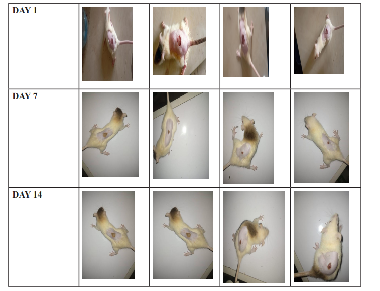
Following wound induction, images were taken across the various test groups to show the rate of wound healing. These images are presented below:

3.3: Fasting Blood Glucose Results
Alloxan was used to induce diabetes in the test animals. Their blood glucose levels were measured before and after induction. These values are represented in Table 2.

3.4 RESULTS OF HISTOLOGY
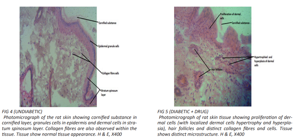
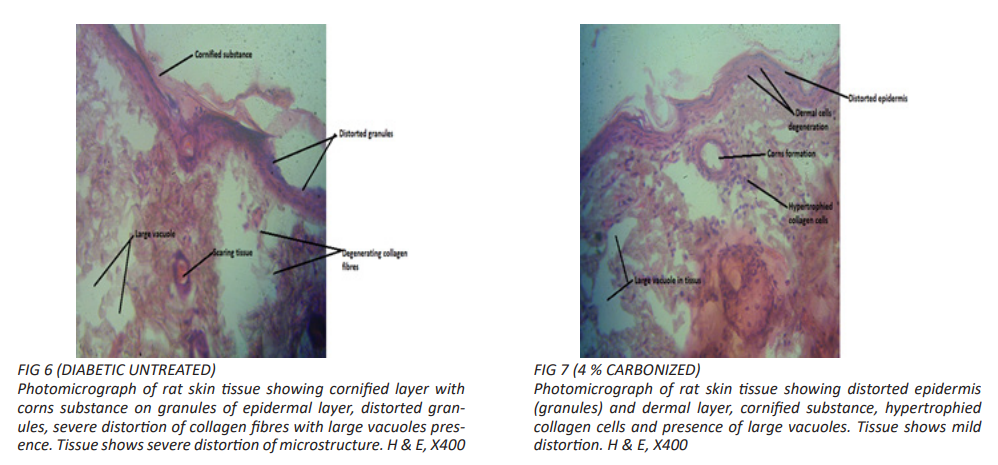
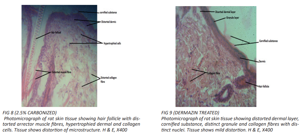
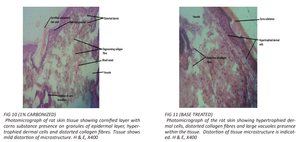
The results of the histological investigations on the wound healing activities of the various test samples and the control groups are presented in Figure 4 – 12 below.

Table 1: Showing physicochemical parameters of the seed pod of Pentaclethra macrophylla Benth.

Figure 1: Wound contraction of different test groups on Days 1, 7 and 14.

Table 2: Table showing the blood sugar readings of the various groups, before induction, after induction, and on Days 7 and 14.

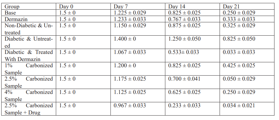
Table 3: Showing the mean and standard error of mean, of the wound contraction in centimeters (cm) on Days 7, 14 and 21.

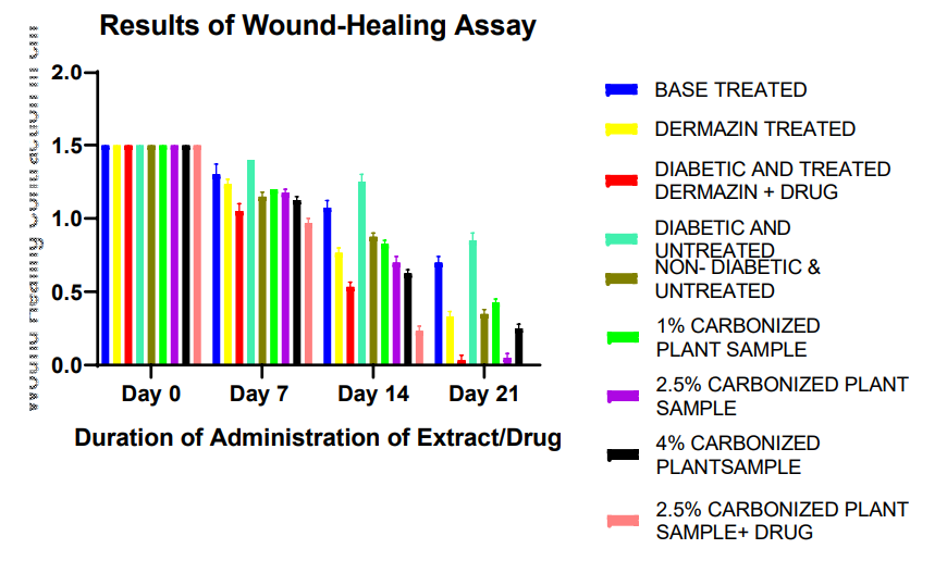
Figure 2: Chart showing wound contraction in centimeters (cm) for control (Based treated, dermazin treated, diabetic and treated with dermazin plus drug, diabetic and untreated, non-diabetic and untreated, and test group (1% carbonized plant sample, 2.5% carbonized plant sample, 4% carbonized plant sample, 2.5% carbonized plant sample plus drug).

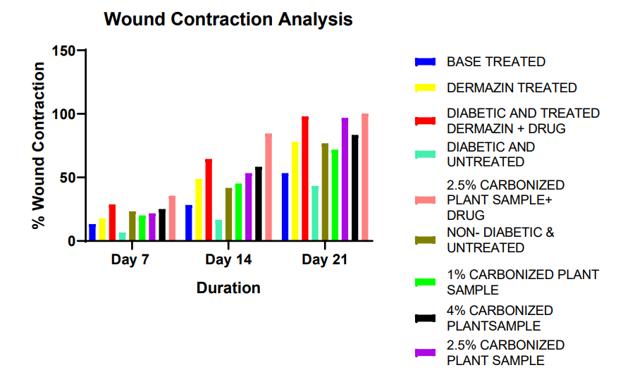
Figure 3: Chart of wound contraction in percentage (%) for control group (Based treated, dermazin treated, diabetic and treated with dermazin plus drug, diabetic and untreated, non-diabetic and untreated and test group (1% carbonized plant sample, 2.5% carbonized plant sample, 4% carbonized plant sample, 2.5% carbonized plant sample plus drug).

Figures 4 & 5

Figures 6 & 7

Figures 8 & 9

Figures 10 & 11

Figures 12: (2.5% CARBONIZED + DRUG) Photomicrograph of rat skin tissue showing distorted epidermal granules, scaring tissue, distinct collagen fibres with distinct nuclei. H & E, X400.

DISCUSSION

Pentaclethra macrophjylla Benth (Fabaceae) is a plant with a long history of traditional use in wound healing [10]. The result of the quality control analysis of the seed pod of Pentaclethra macrophylla is shown in Table 1. The acid-insoluble ash and water-soluble ash values are 1.0% and 2.5% respectively. High acid-insoluble ash values may indicate contamination or poor quality of the sample, affecting its suitability for various applications, while excessive water-soluble ash may suggest the presence of unwanted salts or substances, which can affect the product's quality, taste, or medicinal properties [19]. The low acid insoluble and water soluble ash values suggest that the sample has fewer non-volatile and inorganic contaminants.

DFU is a major complication of diabetes that results in increased hospitalization time, increased health costs, and even death [20].

The wound healing process is a myriad of events. These include homeostasis; the recruitment of platelets and other clotting factors in response to a leaking vessel, inflammation; which involves the invasion of macrophages and neutrophils to clear up any pathogens at the injury site, proliferation; which involves the keratinocytes and matrix deposition to enhance wound closure and remodeling; where collagen is remodeled to finish up the wound healing process [21]. The aim of this study was to compare the different strengths of the carbonized plant extract by evaluating their effectiveness in diabetic wound healing.

From the results obtained, the 2.5% + drug group gave the highest percentage of wound contraction. This was followed by the dermazin + drug group, which is not surprising as the drug; glibenclamide is a sulfonylurea that increases insulin sensitivity of cells. When comparing the Dermazin + drug and the 2.5% + drug groups, they both gave similar percentage wound contraction rates. However, comparing the 1%, 2.5%, and 4% carbonized drug groups, the 2.5% gave a comparable percentage of wound healing as the 4% group. While a comparison of the percentage contraction of the dermazin group to that of the 2.5 and 4% groups, revealed that these two groups demonstrated superior wound healing activities; pointing towards the powerful wound healing effects of the plant. When comparing the results of the 2.5% and 4% groups, it was bserved that the 4% group had a slightly higher wound healing rate from day 0 to 14, which was not statistically significant. But on day 21, the 2.5% treated group showed slightly better results; which were not statistically significant than that of the 4%; suggesting that the 2.5% extract would be the optimal dose for wound healing.

A comparison of the wound healing activities of the extracts and the positive standard, showed that the plant extracts gave a better wound healing profile. Despite the decline in the pursuit of natural products as potential drug leads, encouraging results, like this obtained by the plant extracts of P. macrophylla, provide hope for the use of natural products in disease treatment.

Conclusion

This study demonstrated the wound healing effect of the carbonized seed pod extracts of Pentaclethra macrophylla, with the 2.5% plant extract showing the optimal wound healing activity. This provides a foundation for further research on the plant and its potential as a therapeutic agent for diabetic foot ulcers.

Conflict of Interest
The authors declare that there is no conflict of interest.

REFERENCES

  1. Cho NH, Shaw JE, Karuranga S, Huang Y, da Rocha Fernandes JD, et al. (2018) IDF Diabetes Atlas: Global estimates of diabetes prevalence for 2017 and projections for 2045. Diabetes Research and Clinical Practice 138: 271-281.
  2. Mieczkowski M, Mrozikiewicz-Rakowska B, Kowara M, Kleibert M, Czupryniak L (2022) The Problem of Wound Healing in Diabetes—From Molecular Pathways to the Design of an Animal Model. International Journal of Molecular Sciences 23(14).
  3. Jiang P, Li Q, Luo Y, Luo F, Che Q, et al. (2023). Current status and progress in research on dressing management for diabetic foot ulcer. Frontiers in Endocrinology 14: 1221705.
  4. Clayton WJr, Elasy TA (2009) A review of the pathophysiology, classification, and treatment of foot ulcers in diabetic patients. Clinical Diabetes.
  5. Brem, Harold, Tomic-Canic, Marjana (2007) Cellular and molecular basis of wound healing in diabetes. The Journal of Clinical Investigation 117(5): 1219-1222.
  6. Armstrong DG, Boulton AJ, Bus SA (2017) Diabetic foot ulcers and their recurrence. The New England Journal of Medicine 376(24): 2367-2375.
  7. Walsh JW, Hoffstad OJ, Sullivan MO, Margolis DJ (2016) Association of diabetic foot ulcer and death in a population-based cohort from the United Kingdom. Diabetic Medicine 33(11): 1493-1498.
  8. Goodridge D, Trepman E, Sloan J, Reid G, Embil J (2005) Quality of life of adults with unhealed and healed diabetic foot ulcers. Foot & Ankle International 26(11): 899-910.
  9. Kim J, Nomkhondorj O, An CY, Choi YC, Cho J (2023) Management of diabetic foot ulcers: A narrative review. Journal of Yeungnam Medical Science 40(4): 335-342.
  10. Soepadmo E (2016) Pentaclethra macrophylla: A traditional wound healing plant. Journal of Ethnopharmacology.
  11. Okorie CC, Oparaocha ENT, Adewunmi CO, Iwalewa EO, Omodara SK (2006) Anti-inflammatory and cytotoxic activities of Pentaclethra macrophylla aqueous extracts in mice. African Journal of Traditional, Complementary and Alternative Medicines, 3(1): 44-53.
  12. Oyejide SO, Nwinyi O, Oniha MI, Olugbuyiro J, Taiwo O (2022) Antibacterial activities of Pentaclethra macrophylla and Syzygium samarangense against opportunistic bacterial pathogens. International Journal of Advances in Life Science and Technology 6(1): 24-30.
  13. Nwagwe OR, Eze AC, Nweke EJ (2024) Exploring the therapeutic potential of Pentaclethra macrophylla in diabetic wound management. Journal of Ethnopharmacology, 295: 115326.
  14. AOAC (1990) Association official Analytical chemists. 15th edn. Wash. Dc, U.S.A.
  15. Kim M (2024) Induction of Diabetes Mellitus Using Alloxan in Sprague Dawley Rats. Cureus 16(6).
  16. Samanta R, Pattnaik AK, Pradhan KK, Mehta BK, Pattanayak SP, et al. (2016) Wound Healing Activity of Silibinin in Mice. Pharmacognosy Research 8(4): 298-302.
  17. Quazi A, Patwekar M, Patwekar F, Mezni A, Ahmad I, (2022) Evaluation of Wound Healing Activity (Excision Wound Model) of Ointment Prepared from Infusion Extract of Polyherbal Tea Bag Formulation in Diabetes-Induced Rats. Evidence-Based Complementary and Alternative Medicine: ECAM.
  18. Slaoui M, Bauchet AL, Fiette L (2017) Tissue sampling and processing for histopathology evaluation. Methods in Molecular Biology pp. 1641.
  19. Chandel HS, Pathak AK, Tailang M (2011) Standardization of some herbal antidiabetic drugs in polyherbal formulation. Pharmacognosy Research 3(1): 49-56.
  20. Boulton AJ, Vileikyte L, Ragnarson-Tennvall G, Apelqvist J (2005) The global burden of diabetic foot disease. Lancet 366: 9498.
  21. Wilkinson HN, Hough LM, Schupp, M, Trowbridge, AS (2020) The role of inflammation in wound healing. Journal of Wound Care 29(10): 542-550.
TOP