Giant pilomatrixoma in a 12 year-old child

Sahar Vanessa Amiri * and Alessandro Venzo

Department of Burns and Plastic Surgery, Rigshospitalet, Copenhagen, Denmark

*Corresponding author

*Sahar Vanessa Amiri, Department of Burns and Plastic Surgery, Rigshospitalet, Copenhagen, Denmark

Abstract

Pilomatrixoma is a benign adnexal tumor characterized by hair matrix differentiation, most commonly observed in children. Clinically, it typically presents as a slow-growing, pink to purplish dermal nodule. Due to its low diagnostic accuracy, pilomatrixoma is frequently misidentified as other types of tumors. This case highlights an atypical presentation of pilomatrixoma in a 12-year-old child and underscores the importance of considering ultrasonography or proceeding directly to surgical excision for definitive diagnosis.

Introduction

Pilomatrixoma is a slow growing benign tumour derived from the matrix cells of the hair follicle, also referred to as Malherbe’s calcifying epithelioma [1]. It commonly presents as an asymptomatic, firm, slow-growing, and mobile nodule, most frequently observed in the head and neck region [2]. This case report illustrates a presentation of pilomatrixoma, reported due to its rarity, variable clinical manifestation, and the need to distinguish it from other soft tissue tumors.

Case
We present a 12-year-old healthy girl with no medical history, who was referred to the department of pediatric oncology under a cancer diagnostic program with a progressively enlarging palpable mass over the right scapular region. The tumour had been growing over a period of two years without associated pain, pruritus, or discharge. The patient’s parents reported an accelerated growth over the past four months. The patient was initially assessed by her general practitioner, who referred her for an ultrasound examination. The ultrasound revealed a hypoechoic, hypervascular solid mass measuring 2.4 × 2.3 × 1.6 centimetres, raising suspicion of malignancy. On physical examination in the hospital, we observed a soft localized tumour. The mass was painless when palpating, mobile and doubled in size, measuring approximately 4 × 4 × 2 cm. No clinical signs of lymphadenopathy or other cutaneous lesions were observed. The tumour did not have any effect on range of motion.  Since ultrasound was in this case nondiagnostic, the physicians decided on needle biopsies. At Rigshospitalet, coarse needle biopsies were performed under general anaesthesia, and histopathological analysis confirmed the diagnosis of pilomatrixoma without evidence of malignancy.

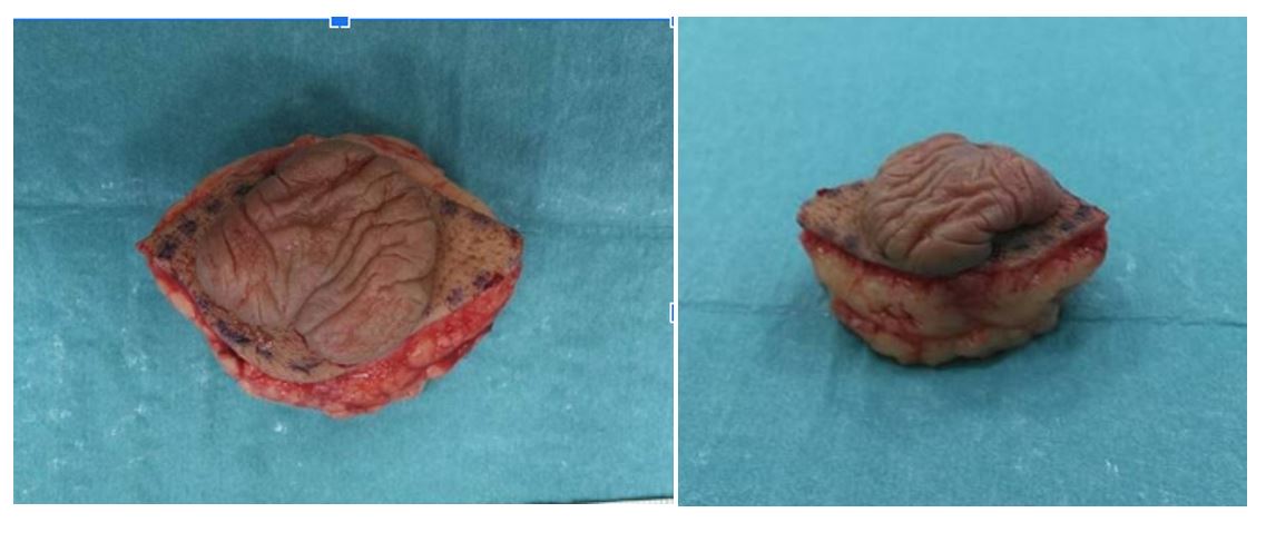
Due to the rapidly progressive growth history the case was discussed in a multidisciplinary team (MDT) conference involving pathologists, paediatric surgeons, thoracic surgeons, paediatric-oncologists, and radiologists. A consensus was reached to proceed with surgical excision. The patient underwent macroscopic radical excision of the tumor under general anesthesia, and the defect was closed with direct closure. Post-operatively, we observed no complications. Histopathological evaluation of the resected specimen confirmed the diagnosis of pilomatrixoma. No follow up was deemed necessary.

Discussion

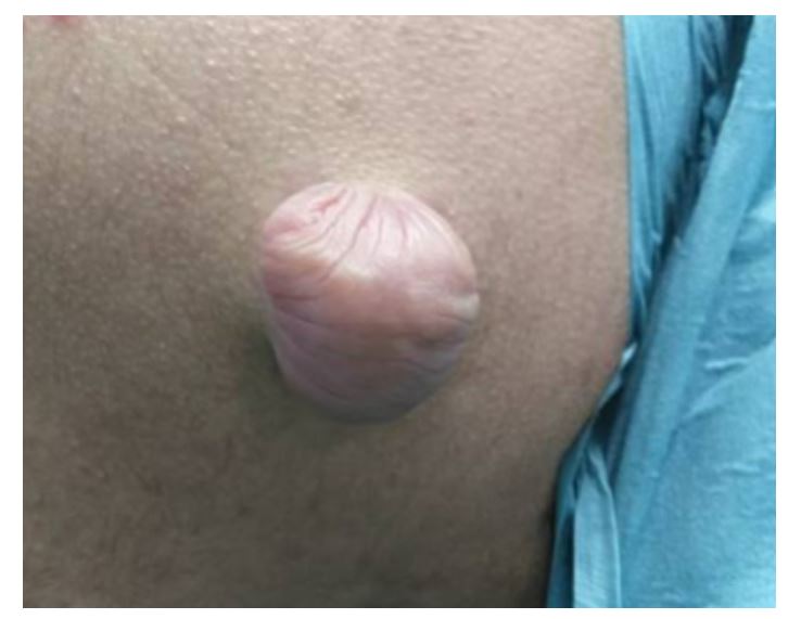
Pilomatrixoma, also known as Malherbe’s calcifying epithelioma, was first described in 1880 by Malherbe and Chenantais [3]. Diagnosing pilomatrixoma can be challenging due to its varied presentation. Histologically, pilomatrixomas presents as a well demarcated tumour, islands of basaloid cells with abrupt keratinization, shadow cells and basophilic cells with normal mitotic activity [4]. While it is predominantly seen in children and young adults, cases have also been reported in adults. Most cases present in the head and neck region, however, around 20% is reported on the trunk [5]. Studies have reported an average size of 1.2 cm [6]. Tumours exceeding four centimetres in size with rapid progression are classified as giant pilomatrixomas [7]. Giant pilomatrixomas can have ulceration, making it clinically difficult to extinguish from squamous cell carcinoma.  Pilomatrixomas often present as asymptomatic nodular lesions. The overlying skin is typically normal but may occasionally exhibit a bluish or reddish discoloration [1]. In this case, the patient had a light nodular element, the skin stretched out, causing part of the lesion to flatten and take on an angular, tent-like shape.

Differential diagnoses include dermoid cysts, neurofibromas, haematoma, basal cell epithelioma, and epidermoid cysts, among others. A rare malignant variant, pilomatrix carcinoma, has also been described. Pilomatrix carcinoma was first reported in 1980, and to date, only 125 cases have been documented in the literature [8,9].  A review on pilomatrix carcinoma from 2017 presented three cases of pilomatrixoma carcinoma in previous scar from benign pilomatrixomas. The majority of reported cases of malignant transformation have been observed in adults, particularly in the 40 to 50 year age range. However, rare cases have also been described in children.

In cases of multiple pilomatrixomas, further evaluation for syndromic associations is warranted, as this condition has been linked to Gardner syndrome and Turner syndrome, as well as others [10].  Additionally, pilomatrixomas have a higher prevalence among patients with myotonic dystrophy, a disorder associated with an increased risk of certain malignancies. There is no well-defined standard treatment protocol for pilomatrixoma; however, complete surgical excision is recommended to ensure total removal of the lesion and to prevent recurrence.

In conclusion, pilomatrixomas are rare and mostly benign tumour, albeit diagnosis is essential in order to exclude malignancy.

Figure 1:Tumour preoperatively.

Figure 2: Tumor shrinking after excision.

Conclusion

This case emphasizes how sarcoidosis can closely mimic malignancy, particularly through blastic bone lesions, hypermetabolic activity on PET imaging, and non-caseating granulomas on biopsy. These findings, often seen in metastatic cancer or lymphoma, highlight the need for careful histological evaluation to avoid misdiagnosis. Recognizing this overlap is essential for accurate diagnosis and appropriate management.

REFERENCES

    1. Jones CD, Ho W, Robertson BF, Gunn E, Morley S (2018) Pilomatrixoma: a comprehensive review of the literature. Am J Dermatopathol 40:631–641.
    2. Zhao A, Kedarisetty S, Arriola AG, Isaacson G (2021) Ear Nose Throat J. 2021:1455613211044778.
    3. RAAB W (1963) Epithelioma of the sebaceous glands. Archiv fur Klinische und Experimentelle Dermatologie 216: 325-333.
    4. Laffargue JA, Stefano PC, Vivoda JL, Yarza ML, Bellelli AG, (2019) Pilomatrixomas in children: Report of 149 cases. A retrospective study at two children's hospitals. Arch Argent Pediatr 117(5): 340-343.
    5. Panda, Sangram K, Sahoo, Pradyumna K, Agarwala, (2023) A systematic review of Pilomatrix carcinoma arising from the previous scar site of Pilomatrixoma. Journal of Cancer Research and Therapeutics 19(5): 1098-1102.
    6. Pirouzmanesh A, Reinisch JF, Gonzalez-Gomez I, Smith EM, Meara JG (2003) Pilomatrixoma: a review of 346 cases. Plast Reconstr Surg 112(7): 1784-1789.
    7. Sabater-Abad J, Matellanes-Palacios M, Bou-Boluda L, Campos-Dana JJ, Alemany-Monraval P, Millán-Parrilla F (2020) Giant pilomatrixoma: a distinctive clinical variant: a new case and review of the literature. Dermatol Online J 26(8).
    8. Jones C, Twoon M, Ho W, Portelli M, Robertson BF, (2018) Pilomatrix carcinoma: 12-year experience and review of the literature. J Cutan Pathol. 45(1):33-38.
    9. Laffargue JA, Stefano PC, Vivoda JL, Yarza ML, Bellelli AG, et al.(2019) Pilomatrixomas in children: Report of 149 cases. A retrospective study at two children's hospitals. Arch Argent Pediatr. 117(5):340-343.
    10. Levy J, Ilsar M, Deckel Y, Maly A, Anteby I, Pe'er J (2008) Eyelid pilomatrixoma: a description of 16 cases and a review of the literature. Surv Ophthalmol 53(5):526-535.
TOP