PRESENTATION OF JUVENILE METACHROMATIC LEUKODYSTROPHY – A RARE CASE REPORT

Niharika Khullar *

Department of Pediatrics, Marengo Asia Hospitals, Faridabad, Haryana, India

*Corresponding author

*Niharika Khullar, Department of Pediatrics, Marengo Asia Hospitals, Faridabad, Haryana, India

BACKGROUND

Metachromatic leukodystrophy (MLD) is the neurometabolic disease caused by deficiency of enzyme arylsulfatase a resulting in deficiency of sulfatide degradation. The responsible gene is arylsulfatase A gene and is inherited in an autosomal recessive manner. MLD is characterized by three clinical subtypes, defined primarily by age at presentation such as late infantile MLD, juvenile MLD, adult onset MLD. Here we report a case of Juvenile form of MLD that was identified by means of typical history, clinical findings, typical MRI brain findings

CASE REPORT

A 15-year-old male child born out of non-consanguineous parents presented with the history of weakness of both upper and lower limb for last one year with progressive impairment and clumsiness of gait for last 6 months; deterioration of school performance was noted for last 6 months.

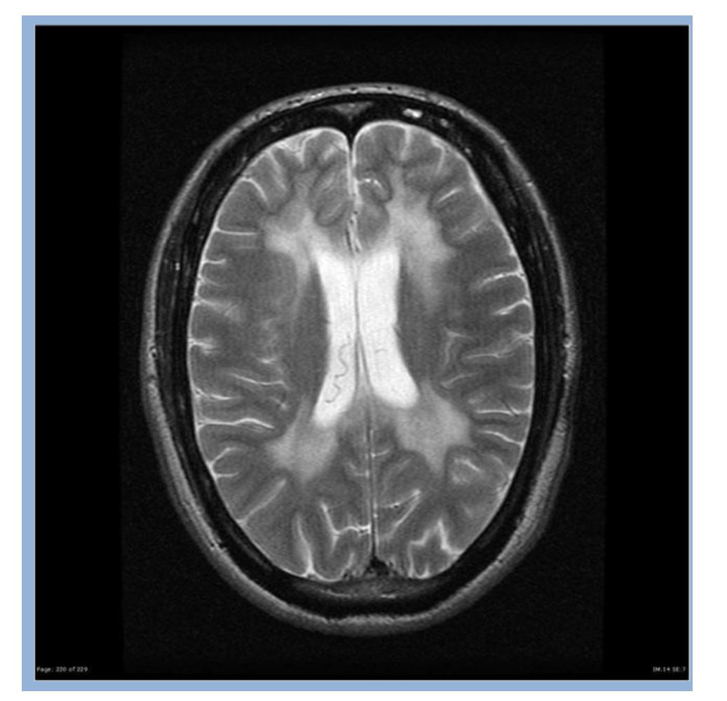
The vision and hearing of the patient were seemed to be normal. His perinatal period was uneventful and developmental milestones were age appropriate prior to this illness. On examination, the child looked apathetic with poor facial expression; he had a somewhat mask like appearance; furthermore, there was no facial dysmorphism and there were no neurocutaneous markers. On examination there is loss of immediate memory, repetition of speech, reduced attention span, hypertonia with exaggerated reflexes, dysdiadokinesia with abnormal gait. MRI showed cerebral cortical atrophy, dilatation of ventricular system, reduced volume of white matter noted in bilateral periventricular region. T2 and FLAIR hyperintense signals are noted in B/L subcortical and deep white matter showing no diffusion restriction.

Discussion

Metachromatic leukodystrophy (MLD) belongs to a family of disorders identified as lysosomal storage diseases. This disorder is characterized by the lysosomal accumulation of sulfated glycolipids, specifically 3-O-sulfogalactosylcontaining glycolipids, as a consequence of defects in the lysosomal hydrolase, arylsulfatase A(ARSA). The major site of 3-O-sulfogalactosyl-containing glycolipids is the myelin sheaths of central and peripheral neurons. Because of this location the clinical manifestations of MLD are predominately neurological in nature. Histopathologically, MLD is characterized by demyelination of central and peripheral nerves. The accumulation of sulfated glycolipids in the lysosomes results in the characteristic metachromatic staining of the tissues, hence the derivation of the name of this disease. A copy of the defective gene from both parents should be transmitted to the offspring to have thedisease. Parents can each have the defective gene and remain as carriers but not have MLD. Children who inherit only one defective gene from one parent will be a carrier, but usually will not develop MLD. When two carriers have a child, there is a 25% chance that the child will get both genes and have MLD. Clinical features of MLD include mental deterioration, hypotonia (low muscle tone), developmental delay, speech abnormalities, loss of mental abilities, blindness, rigidity, convulsions, impaired swallowing, paralysis, dementia, impaired school performance, ataxia, tremors, seizures.

Figure 1

Conclusion

This case emphasizes how sarcoidosis can closely mimic malignancy, particularly through blastic bone lesions, hypermetabolic activity on PET imaging, and non-caseating granulomas on biopsy. These findings, often seen in metastatic cancer or lymphoma, highlight the need for careful histological evaluation to avoid misdiagnosis. Recognizing this overlap is essential for accurate diagnosis and appropriate management.

REFERENCES

TOP