Symptomatic Non-stenosed Carotid Artery causing progressive White Matter Disease Receiving Delayed Intervention

Roberta Potamianos1,2*, Michael Waters1,2 and Jim Jannes1,2

1The University of Adelaide
2Central Adelaide Local Health Network

*Corresponding author

*Roberta Potamianos, The University of Adelaide, Central Adelaide Local Health Network, 15 Stevenson Street, Nailsworth, 5058, SA

Abstract

This case presents an example of severe, progressive white matter disease, secondary to carotid atherosclerosis and small vessel disease. It raises two issues: firstly whether carotid digital subtraction angiography, the gold standard evaluation for carotid stenosis should be offered earlier in the setting of recurrent insults to the same vascular territory and secondly, whether the threshold for intervention should be lower for Symptomatic Non-Stenotic Carotid Artery Disease (SyNC).

Keywords: Stroke; Symptomatic non-Stenotic Carotids - ‘SyNC’; Endarterectomy; Carotid stent

Case report

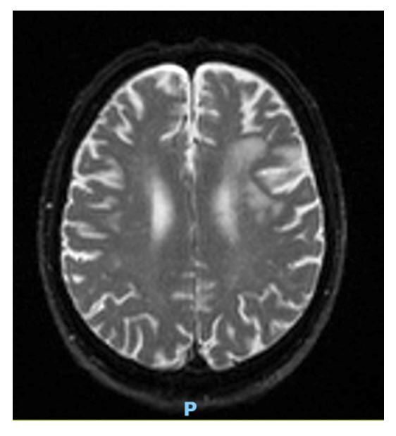
We report a case of an 84-year-old male who presented with acute on chronic neurological deterioration in the setting of a symptomatic atherosclerotic carotid artery. The patient was first admitted under the stroke unit in 2021, when he presented with acute onset right upper limb weakness on a background of hypertension, type 2 insulin-dependent diabetes, hypercholesterolaemia and migraines. MRI of the brain revealed multi-focal acute on chronic left middle cerebral artery (MCA) strokes (Figure 1, LEFT). Non-invasive imaging identified a left internal carotid artery (ICA) stenosis of less than 50% and therefore no surgical intervention was pursued, despite the stroke having been accredited to this left ICA atherosclerosis. Management therefore entailed antiplatelet therapy and optimisation of vascular risk factors. During clinic follow up in the subsequent three years, the patient presented with mild but progressive cognitive impairment which corresponded to progressive left hemispheric subcortical white matter pathology on serial CT brain imaging. These changes, were restricted to the left hemisphere, within the internal and external watershed zones.

In 2024, the patient was readmitted with a two-week history of fluctuating expressive dysphasia. Episodes were observed by family to last for 10 - 15 minutes with spontaneous resolution. Examination revealed that he was hemodynamically stable with a systolic blood pressure of 140mmHg, and in sinus rhythm on 12 lead electrocardiograms. He demonstrated subtle conductive and expressive speech deficits, but no nominal deficits. There were no motor, or sensory deficits and visual fields were normal on bedside testing. The patient was oriented however cognitive testing revealed deficits with recall, loss of short-term memory and tangential speech.

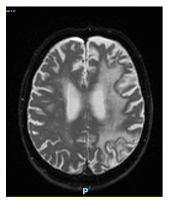
Repeat neuroimaging with MRI brain demonstrated an acute infarct at the left pre-central gyrus on a background of multiple left hemispheric infarcts involving the internal and external watershed zones (Figure 2). The white matter changes had significantly progressed compared to those from the index stroke admission in 2021. Repeat CT angiogram demonstrated mild stenosis of the intracranial left MCA and increased stenosis of the left ICA, now reported at 50-69%, and confirmed on carotid ultrasound.

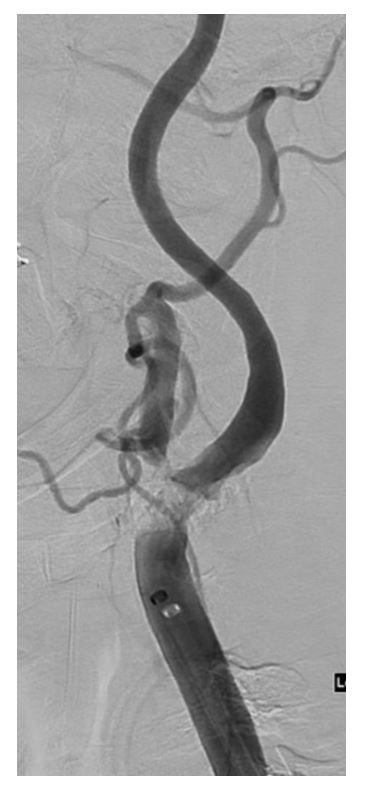
Following multi-disciplinary discussions, and given the evidence of increased left ICA stenosis, the consensus was for intervention. Given the patient’s age and comorbidities, ICA stenting was chosen over endarterectomy. During the intra-operative angiogram, a high-grade left ICA stenosis was identified (Figure 2), higher grade than previously suggested by ultrasound or CT angiogram. It is hoped that improved flow through the patient’s left ICA post-stenting will prevent further cerebral insults and white matter changes.

Figure 1: MRI Auto-Diff TRACEW revealing subacute white matter changes in L MCA territory from 4 years prior (2021 imaging).

Figure 2: MRI – Auto-DIFF-TRACEW  October 2024 at time of presentation demonstrating marked confluent L hemispheric white matter changes.

Figure 3: Digital Subtraction Angiography of Left ICA pre-stenting demonstrating heavily calcific, high grade stenosis.

Figure 4: Digital Subtraction Angiography of Left ICA post-stenting.

Discussion

The threshold for intervening on carotid stenoses remains controversial and the subject of ongoing research, given the risks associated with both carotid endarterectomy and ICA stenting. Both interventions are preventative against ipsilateral stroke in a symptomatic stenosed carotid artery [1,2], and are indicated if greater than 50% luminal stenosis can be demonstrated with clearly associated symptoms [3]. However, the randomised trials supporting these recommendations, specifically NASCET [4] and ECST [5], were published decades ago, and surgical interventions and medical therapies have all improved since then.

At index presentation in 2021, our patient’s carotid stenosis was assessed to be < 50% and therefore fell into a category now recognised as a Symptomatic Non-Stenosed Carotid -‘SyNC.’ A SyNC typically causes subclinical micro-emboli producing subclinical brain infarcts that cumulatively result in cognitive impairment over time [6]. Recent trials are beginning to address whether intervention can be beneficial in these cases [7]. Specifically, whether analysis of alternative biomarkers beyond only luminal stenosis, can help predict which patients would benefit from carotid artery intervention at a lower degree of stenosis to prevent carotid artery plaque related infarctions [8,9].

When our patient re-presented in 2024 with progressive disease as seen on MRI (Figure 2), intra-procedural digital subtraction angiography (DSA) discovered a higher-grade stenosis than identified on non-invasive imaging. Intra-arterial DSA is not recommended as a first line investigation for patients with carotid atherosclerotic disease given the risk of angiogram related stroke [10]. Instead, non-invasive imaging including carotid ultrasound and CT angiogram, is preferred for initial assessment as were used in our case. However, these alternatives, are less sensitive at identifying carotid stenoses, especially at 50-69% stenosis [11,12]. Our case illustrates the limitations of reliance on these modalities and demonstrates that recurrent symptoms and white matter changes in the same vascular territory should trigger investigation with the gold standard test, digital subtraction angiography.

Acknowledgements: Nil

Disclosures: Nil

Conclusion

Although most cases of chronic ITP respond to single agents at relapse, some cases with refractory ITP, require multiple different combinations of all available agents. In the above-mentioned patient, relapse in July 2024 was quite refractory, and multiple rescue agents were used together including Daratumumab (off-label) for several weeks before a response was noted. ITP is in remission presently with oral Fostamatinib. Continued research in this field is needed to develop new agents for treatment in these refractory ITP cases.

Declarations
Consent for Case Report: Written informed consent was obtained from patient to publish this case report in a journal.
Financial Support and Conflict of Interest: None declared

REFERENCES

TOP